Overview Topics

Summary of Scientific Literature

Complement

The complement system is over 20 proteins commonly found in plasma that are part of the so-called innate immune system. These can be traced back in evolution to the most primitive immune defense mechanisms and to the "first line of defense". The two main parts of this network include the activation route (classical pathway) and an amplification loop (the alternative pathway). According to P McGeer, complement is one of the significant factors in neurotoxicity in the AD brain and often found in markedly elevated levels in post-mortem AD brain samples.

The three main actions of the complement protein cascade include opsonization (marking of target invading cells, bacteria, or debris for elimination by phagocytotic cells)[C1q, C3b], anaphylacsis (the activation of other inflammatory signals, including cytokines) [ C4a,C3a, C5a], and formation of the membrane attack complex which leads to the lysis of cells directly. Each of these aspects is regulated in the healthy individual, where checks and balances prevent runaway undesired reactions. For example, inhibitors of the activation of C1 in solution and on cell surfaces and substances that protect cells from complement injury are known. In states of chronic inflammation, including neuroinflammation, such checks and balances fail.

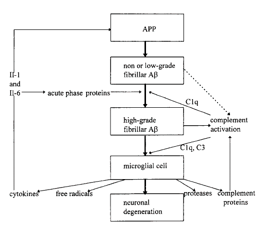
An excellent summary of the interactions of complement, Amyloid beta, and microglia is given by Eikenbloom and Veerhuis (1996) [see link], who posit that early complement factors link the Amyloid Beta deposition to further dramatic inflammatory events. According to the authors, Amyloid Beta is known to bind C1 and to activate the classical complement pathway. It is believed that glial cells are the most likely producers of complement in the brain, and that this production is enhanced by injury. The regulator of C1, called C1-Inh is the only factor known to control C1 activation, and is needed in ratio of 2.5 times the C1 concentration to prevent spontaneous auto-catalysis. C1q complement factor can accelerate Amyloid Beta fibrillogenesis, and the C1q, C3, and C4 that attach to the A-beta deposits activate microglia to express receptors CR3 and CR4.

According to Sayah et al (1999) the optimal concentration of the anaphylatoxin-type complement factors for chemotaxis of cells such as monocytes (e.g. microglia) is 10-8M.

There is inconclusive evidence that points to the presence of the membrane attack complex (MAC) and speculation that this is associated with death of neurons in the cortex in AD patients. Inhibitors of this MAC include CD59, clusterin and vitronectin.

The following diagram, from Eikenbloom and Veerhuis (1996) summarizes the complement-mediated interactions:

Properties of Complement Proteins

The following table, based on Chapter 10 of Neil Cooper (1987) Basic Clinical Immunology, describes the molecular weights and plasma concentrations of various complement factors.

Class Type Molecular
Weight
Typical Plasma
Conc
mu gm/ml
C1 C1q 400 kD 70
C1 C1r 190 kD 34
C1 C1s 87 kD 31
C1-Inh 105 kD 180
MAC C3 185 kD 1300
MAC C5 191 kD 70
MAC C6 120 kD 64
MAC C7 110 kD 56
MAC C8 151 kD 55
MAC C9 71 kD 59

Note: C1={C1q-C1r-C1s} is a Calcium dependent macromolecule, which will associate or dissociate, depending on conditions. C3 forms C3a which is the biologically potent form.

According to Webster (Washington Neuroinflammation Meeting, 2000) C1q inhibits the uptake of A-beta alone, and enhances the uptake of A-beta -antibody complex. The concentration of C1q at which effect is seen is given by the following table:

C1q
Concentration
A-Beta Concentration
for observed effect
muM
1 mu M115 mu M
8 nM0.8 mu M
See Complement References